


Formation of the Early Endosome.

Early endosomes have lowered pH (5.9-6) and this can release the receptor and ligand. The receptor may be recycled to the surface by vesicles that bud from the endosome and then target the plasma membrane. After these recycling vesicles fuse with the plasma membrane, the receptor is returned to the cell surface for further binding and activity. Then, the early endosome converts to a late endosome.
What happen to each receptor in the endosome?

The exact fate of the receptor in the membrane appears to vary with the cell. It can also be degraded. However, some receptors move to the Golgi complex to be added back to membranes in the Trans Golgi region. This would recycle the receptor. This process is similar to the process by which lysosomal enzyme receptors are recycled. In many cases, the receptor is sent back to the plasma membrane after a transport vesicle buds from the endosome. This event is shown in the above photograph. Willingham and Pastan used ferritin labeled antibodies to the extracellular domain of the receptor to follow recycling (transferrin receptor). This photograph was taken from Endocytosis, Edited by Ira Pastan and Mark C. Willingham, Plenum Press, N.Y., 1985
To summarize, markers for early endosomes include rab5-GDP, and pH around 6.0.
Formation of a late endosome
As stated in the above paragraphs, in cases where the receptor is recycled from a budding endosome, the endosome itself is called an "early endosome". In other words, the pH has dropped just enough to allow the ligand to drop off, however, the receptor is not degraded. This keeps it intact so it can be recycled back to the membrane. This is illustrated in the following cartoon.


To understand the formation of late endosomes, you also need to understand sorting in the Golgi complex. Proteins destined for secretion are sorted and packaged into vesicles (see cartoon). However lysosomal enzymes are bound to mannose 6 phosphate receptors and thereby sorted in the Trans Golgi region into vesicles destined for transport to endosomes or lysosomes. Their sorting signal is rab7-GDP. The following cartoon shows the sequestration of lysosomal enzymes in green. The vesicles destined for the Golgi complex are also clathrin coated.

Late endosomes are formed as the pH continues to drop to 5-6.0 thanks to hydrogen pumps on the membrane. The clathrin coated vesicles from the Trans Golgi Network carry digestive enzymes to the late endosome and fuse with these structures, releasing their contents. The lower pH activates these enzymes. The late endosome thus becomes a degradative body and also acquires the marker for mannose 6 phosphate receptor “MPR+”, which carried the enzymes from the Golgi complex. It changes its rab surface marker to rab7-GTP, probably to accommodate the new targeting vesicles with which it will fuse. This means that the late endosome can be identified by the presence of the rab7. The following slide summarizes the key steps in communication from the Golgi Complex.


As summarized above, late endosomes include multivesicular bodies and contain whorls or vesicles of membranes inside. They also contain an unusual lipid which has become another marker for this stage. The lipid is called lysobisphosphatidic acid (LBPA) which has a larger head group than tail. Its structure enables it to be inserted into highly curved membranes, like the membrane whorls. It is believed that this allows retention and binding of specific molecules in the whorls by lipid-protein; lipid-lipid interactions. One type of molecule is cholesterol and it is believed that this is an important site for cholesterol accumulation.
For more information about the role of LBPA, see: Kobayashi, T, Beuchat, M.H, Lindsay, M, Frias S, Palmiter, R.D., Sakuraba, H, Parton, R.G, and Gruenberg, J. Late endosomal membranes rich in lysobisphosphatidic acid regulate cholesterol transport. Nature Cell Biology 1: 113-118.
Late endosomes function to degrade many proteins and lipids. They also are responsible for returning the MPR receptors back to the Trans Golgi network. They recycle these by budding off membranes that carry back the receptors and target the Trans Golgi membranes for fusion. After fusion, the MRP receptors are available to capture and sort new degradative enzymes for future trafficking to the late endosomes.
To summarize, markers for late endosomes include:
- rab7-GDP
- LBPA
- MPR+
Late endosomes fuse with lysosomes.
Finally, late endosomes may not be able to digest all the material. Therefore, the next step is a fusion of late endosomes and lysosomes creating a hybrid organelle. Residual heavily glycosylated lysosomal associated membrane proteins (LAMPs) may thus be transmitted to lysosomes. LAMPs then become a marker for a late endosome or a lysosome. Since lysosomes do not have MPR receptors (they have all been sent to the Golgi), one could distinguish the lysosome and the late endosome on the basis of labeling for MPR. Thus, fusion begins after the MPR have been sent back to the Trans Golgi.

The steps involved in forming late endosomes and lysosomes are drawn in this cartoon. Note that lysosomes continue to communicate with late endosomes and may deliver important material back to this group of organelles. Lysosomes are considered the end product of endocytosis. Thus, lysosomes do not communicate directly with the Trans Golgi (and hence the plasma membrane). However, they could communicate with upstream structures by way of the retrograde transport to the late endosome.

To summarize, markers for lysosomes include:
- LAMP+
- acid hydrolases
- MPR negative
- NPC1 (in normal cells)

Clinical significance of these cellular domains.
Receptor-mediated uptake of LDL receptors: a model for studies of trafficking and defects.

Cholesterol bound to Low density lipoproteins (LDL) is taken up by cells so that cholesterol can be used in construction of membranes, etc. In this case the receptor is recycled and the ligand (LDL-cholesterol) is metabolized so the free cholesterol can be released and used by the cell. We have already looked at the mutation that caused no uptake of LDL receptors. In this section, we will look at how cholesterol could get “stuck in traffic” in the late endosome (see above cartoon). We will look at these diseases to learn more about the importance of these domains.
An important disorder in trafficking is Nieman-Pick C, which causes a build up of cholesterol inside late endosomes. Recent studies point to an important protein involved in cholesterol efflux from the late endosomes, called NPC1. This protein has a putative steroid binding domain. It may be involved in transport of cholesterol to the Trans Golgi Network and then to plasma membranes. Hence it may be important in the addition of cholesterol to membranes where it is needed. It may also be a sensor for sites of accumulated cholesterol.
It got its name from a disease, which is caused by an autosomal recessive mutated gene in which this NPC1 protein is not normal. This is called Niemann Pick type C disorder. The mutation results in an accumulation of cholesterol in late endosomes which also expand, filling with whorls of membranes.
Where is NPC1 protein normally found? Studies by Neufeld et al (J Biol Chem. 274: 9627-9635 (1999) have shown however that NPC1 protein in normal fibroblasts is in the lysosomes (identified by their LAMP marker and absence of MPR). Yet, this same compartment did not contain large amounts of LDL-cholesterol. Thus, Neufeld et al suggested that lysosomes send the NPC1 back to late endosomes by retrograde traffic (see above cartoon). Perhaps this is stimulated by the concentration of cholesterol in the late endosomes. As Mukherjee and Maxfield ( Cholesterol, Stuck in Traffic, Nature Cell Biology 1: E37-E38) point out, this route would then give direct access to the Trans Golgi network to allow trafficking of cholesterol to sites where the cholesterol is needed. Lysosomes don't have routes to these sites, so they may have to communicate to the rest of the cell via late endosomes.
Where is cholesterol needed? Wherever there is a need for more structural stabilization of membranes, for example wherever there are coated pits, particularly clathrin coated pits.
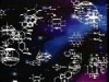
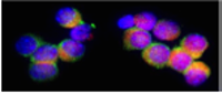
Kobayashi et al studied fibroblasts from Nieman Pick Type C disorder (NPC) and found that not only was cholesterol stuck in the late endosome, mannose 6 phosphate receptors (MPR) are also stuck in this part of the trafficking pathway. Immunolabeling for MPR and CD63 (a marker for late endosomes) showed normal distribution in late endosomes and Golgi regions in normal cells as well as in cells from another disease state, Tay Sach's. However, cells from patients with NPC had cholesterol and MPR almost exclusively in late endosomes. [Kobayashi, T, Beuchat M-H, Lindsay, M, Frias S, Richard D. Palmiter, H Sakuraba, R.G. Parton, and J. Gruenberg, late endosomal membranes rich in lysobisphosphatidic acid regulate cholesterol transport. Nature Cell Biology 1: 113-116 (1999).]
These studies continued by using a drug (U18666A) to mimic the disease state and they found that they could cause both an accumulation of the cholesterol in late endosomes, and also a block in the transport of MPR back to the Golgi complex. Hence, MPRwas detected exclusively with structures labeled for late endosome markers including LBPA and rab7.
What is the significance of these studies? The NPC1 protein may function as a cholesterol sensor/binding protein. An accumulation of cholesterol in the late endosomal compartment may stimulate retrograde transport from lysosomes to endosomes bringing the NPC1 protein to the endosomes. See the pathway and the blocking mechanisms in the above cartoon. There, the cholesterol can be sorted and further distributed to membranes throughout the cell, as needed. If the NPC1 protein is not functional, then cholesterol accumulates in the membrane whorls seen the late endosomes. This appears to block retrograde transport from the endosome to the Golgi complex, as evidence by the accumulation of the MPR. Hence, not only is cholesterol "stuck in traffic", so is the mannose 6 phosphate receptor (MPR). It appears to be stuck as a consequence of the cholesterol accumulation. There is too little known about the return trip to the Golgi complex. However, stay tuned for future information about the role of microtubules and associated motor proteins.
Mukherjee and Maxfield (Nature, Cell Biology, 1: E37-E38 (1999) speculate that perhaps the accumulation of cholesterol in the late endosome makes the membranes less elastic and thus, the return vesicles to the Trans Golgi Network cannot bud and form. There may be broader effects on trafficking as well. Return to the page on membrane architecture for more information about the role of cholesterol in membrane structure and function.
The mechanisms behind the disease are summarized in the following slides.


The two remaining diseases focus on the absence of acid sphingomyelinase (ASM) in lysosomes, which are needed for lipid breakdown. Mutations in ASM cause build up of lipids, which will eventually kill the cells and damage its organ.


For more information, contact:
Gwen Childs, Ph.D.,FAAA
Professor and Chair
Department of Neurobiology and Developmental Sciences
University of Arkansas for Medical Sciences
Little Rock, AR 72205
For questions, contact this email address:
