


An EM view of membranes via freeze fracture/freeze etch .
The freeze-fracture/freeze etch technique starts with rapid freezing of a cell. Then the frozen cells are cleaved along a fracture plane. This fracture plane is inbetween the leaflets of the lipid bilayer , as shown by this cartoon. The two fractured sections are then coated with heavy metal (etched) and a replica is made of their surfaces. This replica is then viewed in an electron microscope. One sees homogeneous regions where there was only the exposed lipid leaflet (Is the exposed surface made of polar or nonpolar groups?
This figure was modified from Bloom and Fawcett, A Textbook of Histology, Chapman and Hall, N.Y., Twelfth Edition, 1994, Figure 1-3.
Consult the section on Membrane Architecture for the answer.)
 In certain areas of the cell,
one also sees protrusions or bumps. These are colored red in the cartoon. Sometimes one
can see structure within the bumps themselves. These are the transmembrane proteins.
In certain areas of the cell,
one also sees protrusions or bumps. These are colored red in the cartoon. Sometimes one
can see structure within the bumps themselves. These are the transmembrane proteins.
The following illustrations will show you some views of specialized regions in the membrane. The organization or structure of the transmembrane proteins can be visualized.
Membrane specializations: Junctions
 The drawing is of a polarized cell. The top is specialized for absorption and the
bottom for transfer of materials to the blood stream. The sides have specialized junctions
that keep the nutrients from entering the space between the cells. For more information
about the thick filaments associated with these junctions, please consult the intermediate
filaments web page.
The drawing is of a polarized cell. The top is specialized for absorption and the
bottom for transfer of materials to the blood stream. The sides have specialized junctions
that keep the nutrients from entering the space between the cells. For more information
about the thick filaments associated with these junctions, please consult the intermediate
filaments web page.
This figure was modified from Bloom and Fawcett, A Textbook of Histology, Chapman and Hall, N.Y., Twelfth Edition, 1994, Figure 2-10
One of these is called a tight junction or "occluding junction" (zonula occludens). This is shown as the top junction in the above drawing. At this site, membrane glycoproteins and associated "glue" bind the cells together like double-sided "strapping tape" The freeze-fracture/freeze etch view of this junction (shown below) illustrates the ridges in the plane of the exposed leaflet. These are the proteins that bind to the proteins from the adjacent cell.
This figure was modified from Bloom and Fawcett, A Textbook of Histology, Chapman and Hall, N.Y., Twelfth Edition, 1994, Figure 2-11.

Gap Junctions
Another type of junction allows communication between cells. This type is called a gap junction. Small molecules or ions can pass through, as we will see by the following figures.

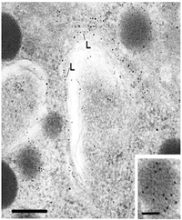
The above freeze-fracture /freeze etch image shows the internal view of the gap
junction on the left. The proteins look like little donuts which reflects the fact
that they are actually a channel. These proteins are "connexon" molecules. The
side facing the cytoplasm (called the P face) is shown in the center panel. The region
looks like aggregated lumps. Finally, the typical electron microscopic view is seen in the
third panel. This shows a thin line between the two plasma membranes indicating a
"gap junction".
This figure was modified from Bloom and Fawcett, A Textbook of Histology, Chapman and Hall, N.Y., Twelfth Edition, 1994, Figure 2-14.
There are several ways to prove the cells are communicating by gap junctions. First,
one can identify the connexon molecules by immunocytochemical labeling. Second, one can
identify the actual junctional complex with freeze-fracture/freeze etch. To see if they
are functional, however, one needs to inject one cell with a dye and watch to see if it is
transferred to another cell.  This cartoon diagrams a view of a gap junction showing
molecules that can freely pass. Ions pass and in this way the cells can be electrically
coupled together. Other small molecules that pass through include cyclic AMP (a second
messenger) and the dye marker fluorescein. This last compound enables the scientist to
study transport throught the gap junction.
This cartoon diagrams a view of a gap junction showing
molecules that can freely pass. Ions pass and in this way the cells can be electrically
coupled together. Other small molecules that pass through include cyclic AMP (a second
messenger) and the dye marker fluorescein. This last compound enables the scientist to
study transport throught the gap junction.
This figure was modified from Bloom and Fawcett, A Textbook of Histology, Chapman and Hall, N.Y., Twelfth Edition, 1994, Figure 2-15.
Return to Menu
Membrane Specializations: Microvilli
The purpose of this final presentation is to introduce a surface specialization that
projects from membranes called the microvillus. It is covered by a plasma membrane and
encloses cytoplasm and microfilaments. Typically microvilli are found in absorptive cells,
whenever there is a need for an increase in surface area.

It is also covered by a glycocalyx which are peripheral glycoproteins that attach themselves to the membrane. It might be used to trap nutrients, protect against toxic subxtances, or adhere to substances needed for uptake. Enzymes used for the cell's function are stored in this region, depending on the cell type.
This figure was modified from Bloom and Fawcett, A Textbook of Histology, Chapman and Hall, N.Y., Twelfth Edition, 1994, Figure 2-17.
The figures to the right show views of microvilli cut transversely.  Note that the microvilli
are lined with the Unit membrane . (top figure) The
core of filaments may allow them to move, although such movement is not as great as that
of cilia or flagella
Note that the microvilli
are lined with the Unit membrane . (top figure) The
core of filaments may allow them to move, although such movement is not as great as that
of cilia or flagella
This figure was modified from Bloom and Fawcett, A Textbook of Histology, Chapman and Hall, N.Y., Twelfth Edition, 1994, Figure 2-18
The lower figure shows a scanning electron micrograph of the luminal surface of the
oviduct. It illustrates one difference between cilia and microvilli. The longer
projections are cilia and the shorter projections are microvilli. For more information
about the internal structure of cilia, consult the Cilia Web page

For more information, contact:
Gwen Childs, Ph.D.,FAAA
Professor and Chair
Department of Neurobiology and Developmental Sciences
University of Arkansas for Medical Sciences
Little Rock, AR 72205
For questions, contact this email address:
