


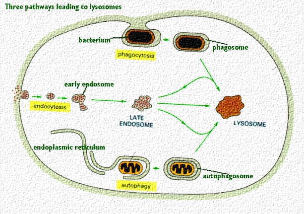
Lysosomes are the cells' garbage disposal system. They degrade the products of ingestion, such as the bacterium that has been taken in by phagocytosis seen in the above cartoon. After the bacterium is enclosed in a vacuole, vesicles containing lysosomal enzymes (sometimes called primary lysosomes) fuse with it. The pH becomes more acidic and this activates the enzymes. The vacuole thus becomes a secondary lysosome and degrades the bacterium.
Lysosomes also degrade worn out organelles such as mitochondria. In this cartoon, a section of rough endoplasmic reticulum wraps itself around a mitochondrion and forms a vacuole. Then, vesicles carrying lysosomal enzymes fuse with the vesicle and the vacuole becomes an active secondary lysosome.
A third function for lysosomes is to handle the products of receptor-mediated endocytosis such as the receptor, ligand and associated membrane. In this case, the early coalescence of vesicles bringing in the receptor and ligand produces an endosome. Then, the introduction of lysosomal enzymes and the lower pH causes release, and degradation of the contents. This can be used for recycling of the receptor and other membrane components. See the Web page on Receptor mediated endocytosis for more information.

Lysosomes carry hydrolases that degade nucleotides, proteins, lipids, phospholipids, and also remove carbohydrate, sulfate, or phosphate groups from molecules. The hydrolases are active at an acid pH which is fortunate because if they leak out of the lysosome, they are not likely to do damage (at pH 7.2) unless the cell has become acidic. A Hydrogen ion ATPase is found in the membrane of lysosomes to acidify the environment.
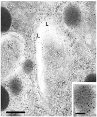
Lysosomal morphology varies with the state of the cell and its degree of degradative
activity. Lysosomes have pieces of membranes, vacuoles, granules and parts of mitochondia
inside. Phagolysosomes may have parts of bacteria or the cell it has injested. This
electron micrograph shows typical secondary lysosomes. They have been detected by
cytochemical labeling for acid phosphatase. This is a good marker for lysosomes. Recall
that it is also used as a marker for the Trans Golgi Cisternae.

The Golgi complex sorts the lysosomal enzyme in the Trans region. It is received from the rough endoplasmic reticulum (RER in this cartoon) in the cis region.
There it has a phosphate radical attached to the mannose residue. This mannose-6 phosphate forms a sorting signal that moves through the cisternae to the trans region where it binds to a specific receptor. After it binds to the receptor, it begins to bud and a "cage" or "coat" made of clathrin forms around the bud (to strengthen it). It moves away to fuse with a developing lysosome (such as the vacuoles seen in the previous figure). This lysosome contains a hydrogen ion pump on its surface. The pump works to acidify the environment inside the lysosome. This removes the phosphate and dissociates the hydrolase from the receptor. The receptor is then recycled back to the Golgi complex
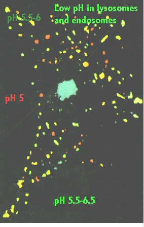
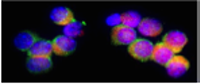
Lysosomes can actually be detected by pH indicator dyes. This photograph shows dyes that indicate different pH's with different colors. The red lysosomes (pH 5.0) are probably typical lysosomes. The blue and green lysosomes are probably endosomes. This change can be detected if you link a ligand to fluorescein. Fluorescein will not fluoresce at pH's lower than 6.0. Therefore, one can follow entry of the receptor-ligand complex and then see the fluorescence disappear as the endosome containing the complex is acidified.
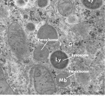
Why Peroxisomes are not like lysosomes.

Peroxisomes are organelles that contain oxidative enzymes, such as D-amino acid oxidase, ureate oxidase, and catalase. They may resemble a lysosome, however, they are not formed in the Golgi complex. Peroxisomes are distinguished by a crystalline structure inside a sac which also contains amorphous gray material. They are self replicating, like the mitochondria. Components accumulate at a given site and they can be assembled into a peroxisome. They may look like storage granules, however, they are not formed in the same way as storage granules. They also enlarge and bud to produce new peroxisomes.
Peroxisomes function to rid the body of toxic substances like hydrogen peroxide, or other metabolites. They are a major site of oxygen utilization and are numerous in the liver where toxic byproducts are going to accumulate.
For more information, contact:
Gwen Childs, Ph.D.,FAAA
Professor and Chair
Department of Neurobiology and Developmental Sciences
University of Arkansas for Medical Sciences
Little Rock, AR 72205
For questions, contact this email address:
