


Answer the question referring to each photograph above the question. The answers are found at the end of the page

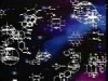
1. Identify the tissue in the above photo.
A) Stratified cuboidal epithelium
B) Simple columnar epithelium
C) Pseudostratified columnar Epithelium
D) Transitional epithelium

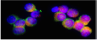
2. Identify the epithelium indicated by the pointer (arrow).
A) cuboidal
B) stratified cuboidal
C) stratified squamous
D) simple squamous
E) transitional

3. Identify the above tissue:
A) Simple columnar epithelium
B) Simple cuboidal epithelium
C) Pseudostratified columnar epithelium
D) stratified columnar epithelium
E) transitional epithelium

4. Identify the tissue indicated by deep pink--red.
A) stratified cuboidal
B) stratified squamous, dry
C) transitional
d) stratified squamous, wet
e) transitional

5. Identify the surface specialization noted by this arrow.
A) Cilia
B) Desmosome
C) Zona adherans
D) Microvilli

6. Name the cytoskeletal elements in this surface specialization.
A) Actin
B) Intermediate filaments
C) Myosin
D) A and C
E) A, B, and C

7. Identify this surface specialization, cut in cross section
A) Cilia
B) Basal Body
C) Centriole
D) Microvilli
E) Desmosome

8. Identify the surface specialization:
A) Zona occludens
B) Hemidesmosomes
C) centrioles
D) Desmosomes

9. Identify the cell type indicated by the arrow:
A) adipocyte
B) fibrocyte
C) Goblet cell
D) myoepthelial cell

10. Identify this type of epithelium
A) Stratified squamous
B) Transitional
C) Stratified cuboidal
D) Stratified columnar
E) Pseudostratified columnar
For more information, contact:
Gwen Childs, Ph.D.,FAAA
Professor and Chair
Department of Neurobiology and Developmental Sciences
University of Arkansas for Medical Sciences
Little Rock, AR 72205
For questions, contact this email address:
Answers: 1C,2D,3B,4B,5D,6E,7A,8D,9C,10B
