
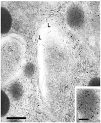
5 nM Gold markers detect LH in the Golgi complex and in a secretory granule.
Immunogold labeling for two or more antigens
This is the favorite electron microscopic immunolabeling protocol in the laboratory of Dr. G. V Childs at the University of Texas Medical Branch . The purpose of this protocol is to allow labeling for two (or more) antigens with sensitive, small protein-A conjugated immunogold markers . This protocol will illustrate our technique for labeling luteinizing hormone (LH), follicle stimulating hormone antigens (FSH) or GH antigens in pituitary cells. In the top Figure, the large gold marks sites of FSH antigens in the smaller storage granules. The small gold marks the site of GH antigens scattered in the largest storage granule on the left. This cell is storing both GH and FSH antigens. For more information about the multipotential somatogonadotrope, see the web page on Growth hormone and gonadotropin co-expression just before ovulation.
The lower figure shows FSH labeling (F) in granules from gonadotropes in estrous rat (top figure) with 10 nM gold markers. The lower figure shows 5 nM gold markers labeling LH (L) in granules in the Golgi Complex.

10 nM gold detects FSH in granules of estrous gonadotrope (Top Figure); 5 nM gold shows LH labeling in granules in the Golgi region.
Materials:
First, make Protein A gold--antibody complex (1:50) stock solutions. We obtain all colloidal gold products from Goldmark Biologicals. Prepare 1% glutaraldehyde in millipore filtered water
Method
- Cut ultrathin sections and place on nickel grids. Etch sections on one side with 0.5% sodium metaperiodate---10 mins. room temperature.
- Immerse/jet-wash 20 times in 0.025M TRIS buffer; blot lightly.
- Block with 5% Bovine serum albumin in 0.025M TRIS buffer---15 mins. room temperature; blot
- Incubate grids in diluted antibody-gold complex (30 microliters/drop) for 2 hours at 33-34 degrees using special tissue culture plates (e.g. 1:3,000 protein A gold-Anti-LH/0.025M TRIS buffer + 5% bovine serum albumin).
- Immerse/wash in 0.025m TRIS buffer & blot against filter paper to dry
- Invert grid to label the other side of the ultrathin sections with the second antibody, etch other side with 0.5% NaIOH—3 min room temperature.
- Immerse/wash in 0.025 M TRIS buffer; blot
- Block with 5% BSA in 0.025M TRIS buffer—15 min room temperature, blot.
- Incubate in the diluted second antibody-gold complex for 2 hours at 33-34 degrees (1:1,500 pAG10-Anti-FSH/0.025M TRIS buffer + 5% BSA)
- Immerse/wash the grids in 0.025M TRIS buffer & dry by blotting on filter paper
- A third antigen can be detected on the first side by 15 nm protein A antibody gold complexes as described for the first, however, the etching step can be omitted. Usually we detect only two antigens at a time.
- Fix the grids in 1% glutaraldehyde---10 mins. room temperature
- Immerse/wash in millipore filtered water & dry by blotting on filter paper
- Counterstain with uranyl acetate & lead citrate. Make solutions one day before use:
- Lead citrate---dissolve 10-40 mg. in 10 ml. Millipore filtered water, add 100 microliters of 10N NaOH, mix solution vigorously
- Uranyl acetate---dissolve 0.8 gm. in 10 ml/ absolute ethanol, mix vigorously; solution must be saturated
- Immerse grid in uranyl acetate drops---5-7 mins. room temperature
- Wash in 25% ethanol---1X by immersion
- Wash in Millipore filtered water---2X by immersion
- Dry grids on filter paper---10 mins. room temperature
- Float on lead citrate drops---5 mins. room temperature
- 0.02N sodium hydroxide---2X by immersion, alternating with millipore filtered water---2X by immersion
- Also dry grids on filter paper
NOTE: lead citrate combines readily with CO2, so keep the dish covered & don't breathe on it. Sodium hydroxide pellets may be used to take up CO2 from air. Make a chamber with sodium hydroxide pellets in one side and a teflon or wax surface for staining the grids on the other side.
Other Protocols:
| Affinity Cytochemistry | |
| In situ hybridization
histochemistry |
For more information, contact:
Gwen Childs, Ph.D.,FAAA
Professor and Chair
Department of Neurobiology and Developmental Sciences
University of Arkansas for Medical Sciences
Little Rock, AR 72205
For questions, contact this email address:
